Introduction: A New Dimension in Ultrasound Imaging
In recent years, the field of ultrasound imaging has witnessed a remarkable advancement that has transformed the way we visualize the human body. The introduction of 3D ultrasound imaging has brought a new dimension to medical diagnostics, offering detailed and lifelike representations of internal structures. The expectant couple chose to have a private pregnancy scan london to cherish their special moments and receive personalized care during this exciting time. In this article, we delve into the fascinating world of 3D ultrasound imaging, exploring its capabilities, applications, and the impact it has on healthcare. Join us as we unravel the extraordinary world of three-dimensional ultrasound imaging.
Understanding 3D Ultrasound: Adding Depth to Diagnosis
Unlike traditional 2D ultrasound, which captures cross-sectional images, 3D ultrasound imaging constructs a three-dimensional representation of the scanned area. By capturing a series of 2D images from different angles, a computer algorithm processes and combines them to create a detailed 3D model. This additional dimension provides healthcare professionals with a more comprehensive view of the internal structures, enabling enhanced visualization and analysis.
Realism and Detail: Bringing Images to Life
One of the key advantages of 3D ultrasound imaging is its ability to bring images to life with exceptional realism and detail. The three-dimensional models allow for a better understanding of the spatial relationships between organs, tissues, and abnormalities. This added depth provides valuable insights for accurate diagnosis, surgical planning, and monitoring of conditions such as fetal development, tumors, and cardiovascular anomalies. The level of realism and detail offered by 3D ultrasound imaging enhances healthcare professionals’ ability to make informed decisions and improve patient outcomes.
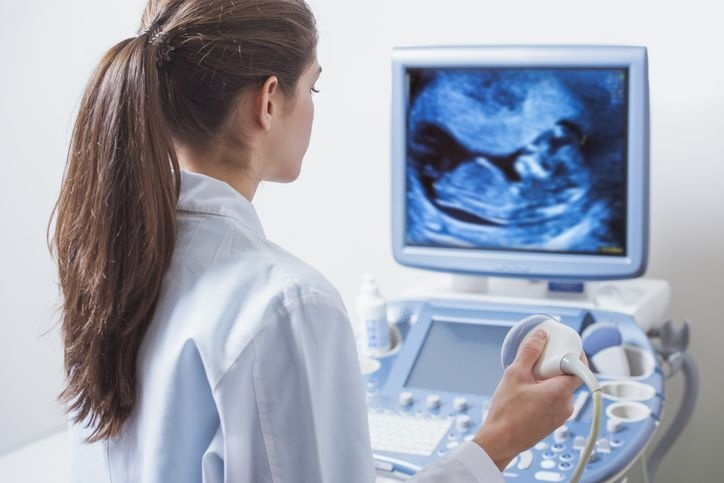
Prenatal Imaging: Capturing Unforgettable Moments
Perhaps one of the most captivating applications of 3D ultrasound imaging is in prenatal care. Expectant parents are now able to see their unborn child in remarkable detail, creating a truly unforgettable experience. The three-dimensional images showcase the baby’s facial features, limbs, and movements, offering a glimpse into their world before birth. This emotional connection between parents and their unborn child can deepen the bond and foster a sense of anticipation and joy. 3D ultrasound imaging has revolutionized prenatal experiences, allowing parents to cherish and share the early stages of their baby’s development.
Complex Anatomy and Pathology: Enhancing Diagnostic Accuracy
In the field of medical diagnostics, 3D ultrasound imaging plays a pivotal role in visualizing complex anatomical structures and identifying abnormalities. It provides a more comprehensive understanding of intricate organ systems, aiding in the detection and characterization of diseases. For instance, in cardiology, 3D ultrasound imaging enables precise assessment of the heart’s chambers, valves, and blood flow patterns. In obstetrics, it helps diagnose fetal abnormalities, such as cleft lip or spina bifida. By enhancing diagnostic accuracy, 3D ultrasound imaging empowers healthcare professionals to provide targeted treatments and interventions.
Surgical Planning and Guidance: Improving Precision
The detailed and realistic nature of 3D ultrasound imaging has proven invaluable in surgical planning and guidance. Surgeons can use pre-operative 3D scans to visualize complex anatomical structures, plan incisions, and simulate procedures. During surgery, real-time 3D ultrasound guidance provides accurate visualization of critical structures, enabling precise navigation and minimizing the risk of complications. This level of precision enhances surgical outcomes and patient safety, particularly in procedures involving delicate areas such as the brain, liver, or heart.
Advancements in Visualization: 4D Ultrasound Imaging
Building upon the foundation of 3D ultrasound imaging, advancements have led to the development of 4D ultrasound imaging, also known as real-time 3D ultrasound. This technology captures a continuous stream of 3D images, creating a dynamic video-like experience. 4D ultrasound imaging has proven particularly valuable in obstetrics, allowing healthcare professionals to observe fetal movements, facial expressions, and even interactions with the environment in real-time. This real-time visualization adds an extra layer of understanding and opens up new possibilities for research and clinical applications.
Conclusion: A New Dimension in Healthcare
In conclusion, 3D ultrasound imaging has revolutionized the field of medical diagnostics by adding a new dimension of depth and realism. Its ability to capture detailed three-dimensional models enhances diagnostic accuracy, improves surgical precision, and provides an emotional connection in prenatal care. The ongoing advancements in ultrasound technology continue to push the boundaries, opening up new avenues for research and clinical applications. With 3D ultrasound imaging, healthcare professionals can uncover hidden details, gain a deeper understanding of the human body, and deliver personalized and effective care to patients.
Leave a Reply